Title III Technology Literacy Challenge Grant
Learning Experience
| LE Title: Anatomical Cardiac Assessment | Author(s): Sara Nicolette & Lori Franklin |
| Grade Level: post secondary(also applicable to high school anatomy & physiology classes) | School Address: Herkimer Co. BOCES 352 Gros Blvd. Herkimer, N.Y. |
| Topic/Subject Area: Human Anatomy & Physiology for Licensed Practical Nurses | School Phone/Fax (315)867-2000 |
LEARNING CONTEXT
Purpose or Focus of Experience
This experience serves as a cumulative application of the knowledge gained in the cardiac unit. It allows the student to incorporate both the declarative and procedural knowledge they gained. This sets the framework for the pathophysiology applications that are taught in their med/surg nursing class.
Connection to Standards
CAREER DEVELOPEMENTAL & OCCUPATIONAL STUDIES
Standard 3b Specialized Health Services
Academic Foundations
Essential Question
The essential question in this unit is two fold. First is to identify anatomical landmarks of the previously dissected heart. Second to explain the identified structures functions in the cardiac cycle.
Content Knowledge
| Declarative | Procedural |
| Cardiac anatomy | Identify anatomical markings of the heart |
| cardiac valve structural variations | Identify normal blood flow patterns |
| left vs. right ventricular structure & rational | identify ventricular walls properly |
| * cardiac conductivity pathways & cardiac muscle responses | * identify atrial & ventricular septum’s |
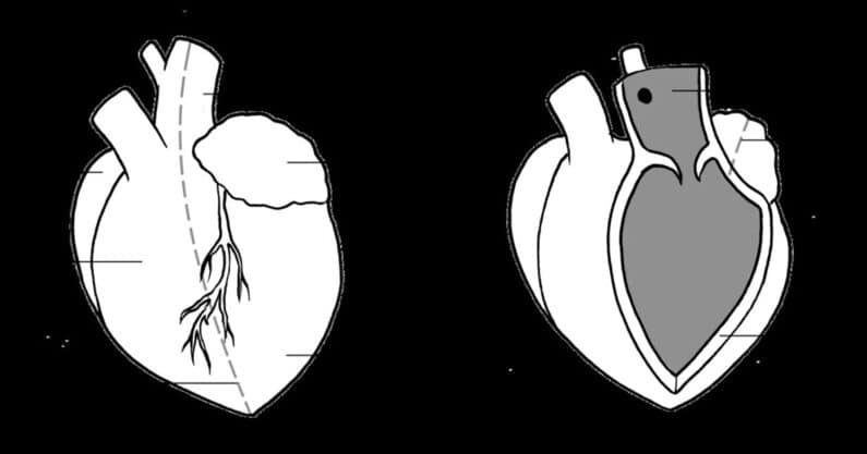
PROCEDURE At the point of this experience the nursing student has participated in several preliminary class exercises. These include required chapter reading on the cardiac system (90 min), lecture on the heart as a muscle, electrical conductor, and the circulatory systems pump (90 min), an interactive class exercise constructing a 6 ft. by 9 ft. walk through floor model of the heart (90 min) and an instructor conducted dissection lad of an actual animal heart & lung (90 min).during the dissection lab digital pictures are takenthese pictures are exported onto a disc for use with power point technologystudents working on groups of two or three are asked to identify the following anatomical sites : right atrium, bicuspid valve, right ventricle, septums, pulmonic valves, left atrium, mitral valve, left ventriclea comparrison on the density of the right & left ventricle is donethe function of each identified anatomical site is reviewed within the groupa written summary of the groups finding is completed by each group participantAll students are reassembled and an instructor lead power point presentation that reviews the individual group findings is done.
INSTRUCTIONAL/ENVIRONMENTAL MODIFICATIONS
The composite class discussion can be modified by transferring the photo prints to overhead transparencies.
TIME REQUIRED
30 to 90 minute prep time depending on method: 90 minute classroom participation time
RESOURCES
- animal heart (easily obtained from a slaughter house or a science supply catalog)
- dissection tray and equipment
- digital camera
- PC Disc for digital picture duplication
- PC ‘s with power point application tools for student
- ability for instructor to give power point presentations
- anatomy & physiology text
ASSESSMENT PLAN
In computer lab groups of 2-3 students will review digital dissection photos
At a 90 % proficiency each group will identify the following anatomical locations: left & right atrium, left & right ventricle, all four heart valves, aorta, cardiac lining, ventricular wall thickness variations, pericardial sac, blood flow patterns
At a 90 % proficiency each group will also explain the function of all identified anatomical locations.
STUDENT WORK
Several digital images prints are enclosed as reference material.
REFLECTION
Use of this technology is a relatively new tool to our classroom. What proved the most helpful was the magnification and clarity that the digital images allowed. Universally the students were enthusiastic about the exercise. It is our intention to duplicate this approach in all future dissection labs. These images will then enable us to have an inventory of dissection exhibits for us at any time as a compliment to a variety of other nursing subjects we teach.

